TuberculosisToday By Pooja Iyer
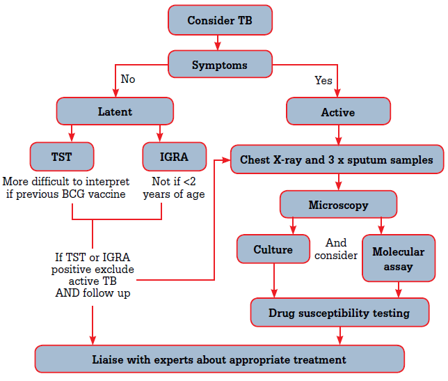
Diagnosis of TB disease includes five important aspects:
1. Medical history of the individual
2. Physical examination by the physician
3. Tests for detecting TB infection
4. Chest X-Ray
5. Bacteriological examination of specimens.
1. MEDICAL HISTORY OF THE INDIVIDUAL:
The clinician will enquire if any symptoms are present and for how long, if there has been any accidental contact with a person infected with TB, and if the individual has been diagnosed with LTBI (Latent Tuberculosis Infection) or TB in the past.
2. PHYSICAL EXAMINATION BY THE PHYSICIAN:
A thorough physical examination may provide crucial information about the condition of the patient, identify and inform the method of diagnosis. But it cannot be used to confirm or rule out TB.
3. TESTS FOR DETECTING TB INFECTION:
The two methods that are widely used for the identification of M. tuberculosis infection are:
-
Mantoux tuberculin skin test (TST);
-
Interferon-gamma release assays (IGRAs)
»QuantiFERON-TB Gold In-Tube test (QFT-GIT)
»T-SPOT®.TB test.
These are differential tests that help to distinguish between people that have been infected with M. tuberculosis with the uninfected individuals. But, a negative result will not rule out the possibility of TB.
4. CHEST X-RAY:
Chest X-Ray can be used to diagnose TB. Abnormalities may indicate pulmonary TB disease.
5. BACTERIOLOGICAL EXAMINATION OF SPECIMENS:
Specimens should be examined in a lab that is specialized in testing for M. tuberculosis. The bacteriological examination has five parts:
• Specimen collection;
• AFB smear culturing;
• Nucleic Acid Amplification;
• Specimen culturing and identification; and
• Drug-susceptibility tests.
-
Specimen Collection Methods for Pulmonary TB Disease
Four specimen collection methods are used for pulmonary TB:
>Coughing
>Induced sputum
>Bronchoscopy
>Gastric aspiration
TB can occur in any part of the body; thus, a wide variety of clinical specimens other than sputum (e.g., urine, cerebrospinal fluid,etc.) can be submitted for examination if extrapulmonary TB disease
is suspected.
-
AFB Smear Culturing
Presence of mycobacteria in a specimen would be evident by the detection of acid-fast bacilli in stained and acid-washed smears that are examined microscopically
Acid-fast staining is done in two ways:
>Carbolfuchsin methods that include the Ziehl-Neelsen and Kinyoun methods (direct microscopy)
>Fluorochrome procedures using auramine-O or auramine-rhodamine dyes (fluorescent microscopy).
-
Nucleic Acid Amplification (NAA)
In this type of test, DNA and RNA segments are amplified and microorganisms are identified in a specimen. It is a quick and reliable method to detect M. tuberculosis bacteria in specimens in a few
hours as compared to the culture method that takes a week or more.
-
Specimen Culturing and Identification
Positive cultures for M. tuberculosis confirm TB; however, if the culture is not positive, clinical signs
and symptoms can also be used to confirm the disease.
Media used for culturing :
>Solid media (Löwenstein–Jensen media)
>Liquid media (MGIT)
-
Drug-Susceptibility Tests
Resistance to the first-line anti-TB drugs: isoniazid, rifampin, ethambutol, and pyrazinamide is checked against the M. tuberculosis isolate obtained from the patient.
Sources:


Let’s Work Together
Get in touch so we can start working together.